What is vocal fold paralysis?
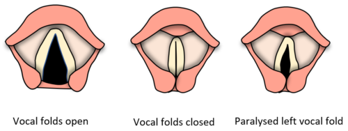
To understand vocal fold paralysis, we should know something about the way voice is produced. Voice is produced when air passes between the closed vocal folds causing the vocal folds to vibrate. The vocal folds are two muscular folds with a soft tissue covering, situated in the voice box just above the windpipe. When we speak, air passes up from the lungs and windpipe between the vocal folds. The vocal folds open in a “V” shape when we breathe, and close when we speak which allows them to vibrate.
 |
The opening and closing movements of the vocal folds is controlled by nerves which follow a complex pathway from the brain, through the neck, into the chest, before making a U-turn and travelling back up through the neck to the voice box.
In vocal fold paralysis there is usually an injury somewhere along the nerve pathway that prevents the opening and closing movements of one or both vocal folds. If one vocal fold is affected this typically results in a weak breathy voice. If both vocal folds are affected, this can cause difficulty breathing.
What problems can vocal fold paralysis create?
When one vocal fold is paralysed, the vocal folds may have difficulty closing. This results in a gap between the vocal folds during voicing. Air leaking through the gap causes the voice to have a weak, breathy quality. Patients may also experience breathlessness on speaking due to excessive air leakage, and difficulty straining and lifting heavy objects. Coughing can be weak or ineffectual and does not seem to clear the throat. Sometimes swallowing is difficult and there may choking spells particularly when drinking liquids.
Paralysis of both vocal folds is rare but can cause life-threatening breathing difficulty. This is because the vocal folds usually become immobilized in the mid-line of the voice box and cannot open into the normal “V” shape for breathing.
What are the causes of vocal fold paralysis?
There are many possible causes of vocal fold paralysis, which are related to injury to the nerve supplying the vocal folds. Causes can include certain surgical procedures in the neck and chest, including thyroid surgery, neck spine surgery, and sometimes surgery on the heart and lungs. Radiotherapy to the head and neck can also occasionally cause injury to the vocal fold nerve. Another cause of paralysis is a tumour in the neck, chest or at the base of the skull, and this is an important reason to ensure that symptoms from vocal fold paralysis are carefully evaluated. People with certain neurological conditions such as stroke or Parkinson disease can also have vocal fold paralysis. There are usually other nerve related symptoms in these conditions, and the vocal fold paralysis is part of that condition. Vocal fold paralysis can also result from certain viral infections. In a proportion of cases referred to as idiopathic there is no identifiable cause.
How is vocal fold paralysis diagnosed?
The diagnosis of vocal fold paralysis is usually suspected from a change in the patient’s voice. Typically, the voice is weak and breathy. There may have been an event such as surgery, trauma, or an infection that preceded onset of symptoms.
A key part of the examination is diagnostic laryngoscopy. This examination is performed in the outpatient clinic and involves passing an endoscope or small flexible camera usually through the nose to visualize the vocal folds. A clear assessment can be made of the structure and movements of the vocal folds, and it is possible to see whether one or both vocal folds is not moving. A strobe light may be used to examine certain aspects of the vocal folds in more detail, such as how well the margins of the vocal folds can meet and close. This part of the examination is known as laryngeal stroboscopy.
Tests may be needed to look for the cause of paralysis. Such tests may include scans of the neck and chest such as an ultrasound scan of the thyroid gland, CT scan or the neck and chest, and / or MRI scan. Sometimes the esophagus or food passage needs to be examined, which can be done easily with a flexible camera passed through nose into the food passage. This examination is known as trans-nasal esophagoscopy and is usually easily accomplished without the need for any sedation, in the outpatient clinic [Read more about trans-nasal esophagoscopy]. Sometimes blood tests can provide additional information.
Laryngeal electromyography is a test that can provide useful information about the function of the vocal fold. In this test a very fine needle is passed through the neck into the muscles of the vocal fold. This enables electrical activity within the muscle to be measured. The test can be help confirm diagnosis of vocal fold paralysis if this remains unclear after diagnostic laryngoscopy. It is also particularly useful to assess the likelihood of recovery of vocal fold movement, which may influence the type to treatment required.
How is vocal fold paralysis treated?
Treatment of vocal fold paralysis is different depending on whether paralysis is one-sided or affects both vocal folds. One-sided paralysis is by far the most common situation.
In one-sided paralysis the first step towards treatment is establishing the cause of paralysis. The cause can give an indication of the likelihood of recovery of the nerve. For example, a nerve that is simply stretched or bruised may be more likely to recover than one that has been cut or damaged by tumour.
The second step is to determine severity of symptoms. Symptoms from vocal fold paralysis range from being very mild to having significant impact on the person’s quality of life, or even creating life-threatening problems such as chest infection due to inability of the vocal folds to close when eating and drinking.
If symptoms are mild and recovery is expected, a wait and see approach is an option. I generally do not advocate prolonged waiting, as this can impair results of subsequent surgery should it become necessary. However waiting for a few weeks, and sometimes months if there are signs of improvement, is not unreasonable. Waiting can be combined with speech therapy to learn how to project and strengthen the voice. Speech therapy can also be helpful for patients with vocal fold paralysis who have swallowing difficulty. If more confirmation is needed on the likelihood of recovery, laryngeal electromyography can be useful.
Sometimes even if recovery is expected, intervention is needed to strengthen the voice. In this situation a temporary minimally invasive procedure may help. This can be achieved by injecting a filler to bulk up the paralysed vocal fold. The bulked-up vocal fold makes better contact with the normal vocal fold, helping voice and swallowing to become stronger. The procedure can be performed either under local anesthesia in the clinic, or under general anesthesia. A filler material is chosen that will gradually reabsorb, allowing the nerve time to undergo recovery.
When a more permanent solution is required, the injection procedure can still give good results, but a non-resorbable filler material should be selected. I generally prefer to perform the injection under general anesthesia for greater accuracy when we are trying to achieve a permanent solution. However, for patients who are not fit enough to undergo general anesthesia, we can still achieve good results with the local anesthetic procedure. An alternative to doing the injection is to place an implant into the voice box through a neck incision. Surgery may be combined with speech therapy to help fine-tune the voice.
Vocal fold injection under general anesthesia for paralysed left vocal fold
Other variations of surgery occasionally used are to adjust the cartilage at the back of the vocal fold or re-innervate the voice box by connecting a healthy nerve from another muscle to the damaged voice box nerve. The newly joined nerve will take some months to regain function, and movements of the paralysed vocal fold may not fully return to normal. The goal of all these procedures is to get the vocal folds to meet in the midline. This enables them to vibrate against each other so that the voice is as strong as possible. As the vocal folds also need to close during swallowing, these surgical procedures can sometimes significantly improve swallowing too.
How is treatment of bilateral (two-sided) vocal fold paralysis different?
When both vocal folds are paralysed, breathing rather than speaking becomes the key concern if the vocal folds become fixed in the midline. Breathing difficulty can range from breathlessness during exertion or exercise, to life-threatening breathlessness even at rest.
Very often patients with severe symptoms will have required a tracheostomy, which is a breathing tube inserted through an incision in the front of the neck into the windpipe. The tracheostomy allows breathing to take place through the tube rather than through the nose and mouth, thus bypassing the obstruction at the vocal folds. Learning how to care properly for the tracheostomy is important.
Surgery can be performed in patients with paralysis of both vocal folds, with the goal of improving breathing and removing the tracheostomy tube. Such surgery is a highly specialised form of micro-laryngeal surgery, which is performed on the vocal folds through the patient’s mouth using special instruments including a laser. [Read more about micro-laryngeal surgery] It is usually not necessary to make any incisions in the neck. Surgery aims to create an opening between the closed vocal folds to allow passage of air for breathing. The difficulty however is that as widening the airway can make the voice worse and create swallowing difficulty. There is a delicate balance between widening the space between the vocal folds and maintaining a good voice and ability to swallow safely. However it is possible to achieve this balance and successfully remove the tracheostomy.
What can we offer at David Lau ENT Centre
Dr Lau is experienced in treating patients with vocal fold paralysis. He will assess you carefully and discuss your treatment options in detail. Dr Lau is recognized as a pioneer of minimally invasive injection methods for outpatient, as well as inpatient treatment of vocal fold paralysis, having introduced out-patient injection medialization to ENT practice in Singapore in 2002. He continues to practice and refine the techniques, and teaches both locally and regionally on the subject.
In addition to minimally invasive techniques, Dr Lau also performs open methods for vocal fold medialization in vocal fold paralysis and has trained in reinnervation techniques in the larynx.
Dr Lau also has experience treating patients with paralysis of both vocal folds and different laser techniques available for this. He has achieved success in decannulating patients with long term tracheostomy for this condition.
He also offers diagnosis with laryngeal electromyography.

